
Near-infrared spectroscopy · Kuopio, Finland
Intraoperative tissue intelligence for knee arthroscopy.
Incight Medical Technologies develops JED Explorer. It uses near-infrared spectroscopy to measure tissue characteristics that visual inspection and probing alone cannot assess, within the familiar arthroscopic workflow, and is being developed to give surgeons objective, quantitative information about the joint when treatment is decided.
JED Explorer is in pre-clinical development. It is not CE marked and is not available for sale.
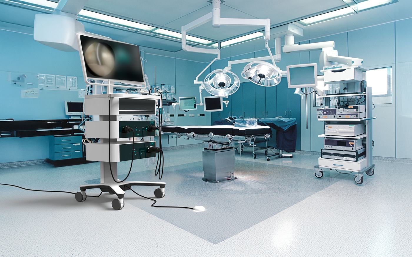
Concept visualisation. JED Explorer has not been used in patients.
Independent evidence
Whether to operate is only the first decision.
In 2026, an international ICRS-FIFA-Aspetar expert panel evaluated six surgical approaches in 16 clinical scenarios of knee cartilage injury in football players, 96 combinations in all. The appropriate treatment depended on factors including lesion location, defect size and whether the damage involved cartilage alone or the underlying bone.
The conclusion was clear: there is no single treatment for every cartilage lesion. The surgical approach needs to be matched to the individual tissue condition and patient.
Papakostas, Kon, Andriolo et al. ICRS-FIFA-Aspetar consensus on the management of knee cartilage injuries in football players, part 2. British Journal of Sports Medicine 60 (2026) 770–779. doi:10.1136/bjsports-2025-110676
JED Explorer is being developed to provide objective, real-time tissue information during arthroscopy to support these treatment decisions.
JED Explorer
The instrument.
JED Explorer pairs a probe shaped like the arthroscopic hook surgeons already use with a compact system cart. Measurement is started with a foot pedal, so both hands stay on the instruments. A working prototype has been tested on animal joints and on human cadaver tissue; clinical validation is the next step.
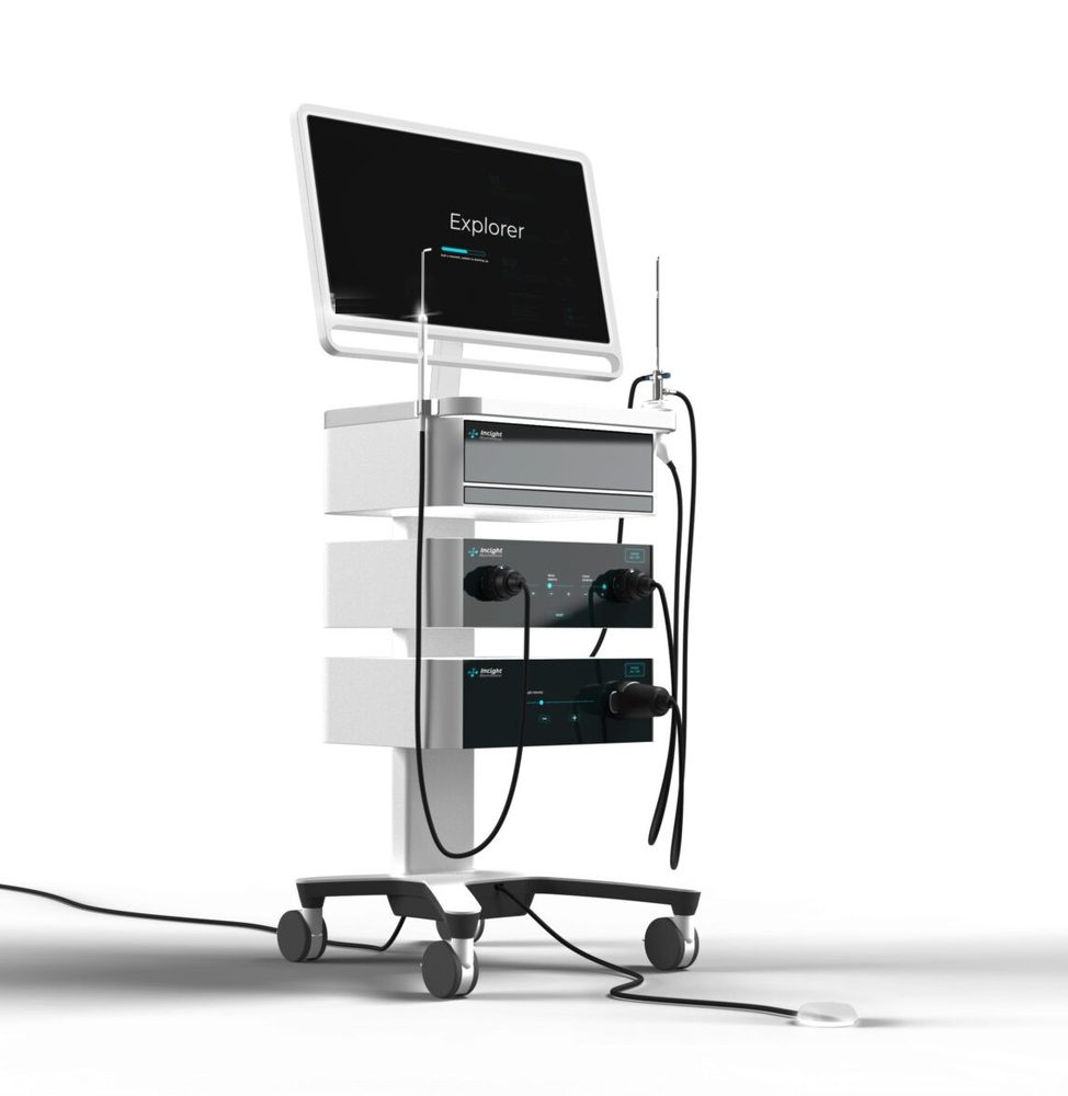
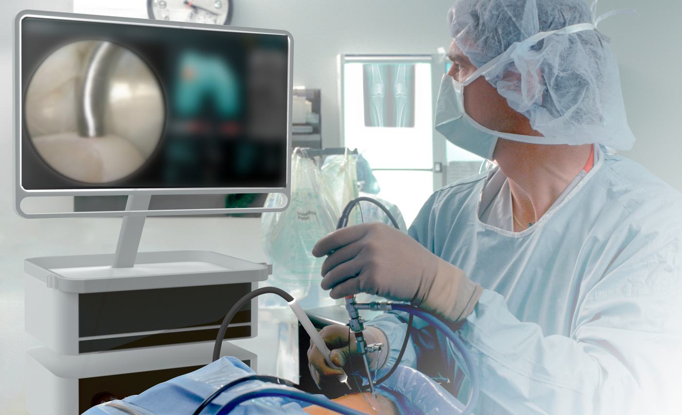
Concept visualisation. JED Explorer has not been used in patients.
Published research
More than a decade in print.
JED Explorer builds on near-infrared spectroscopy research our founders have published since 2013, led by co-founder Isaac Afara and developed at the Biomedical Spectroscopy Laboratory of the University of Eastern Finland.
Articular cartilage
Estimated with near-infrared light in these studiesThickness, and how stiff the cartilage is under the quick, rhythmic and sustained loads of jumping, walking and standing (instantaneous, dynamic and equilibrium moduli).
2013Application of near infrared (NIR) spectroscopy for determining the thickness of articular cartilage
97 bovine cartilage specimens, in the laboratory.
Compared near-infrared spectra with mechanically measured cartilage thickness. The most predictive spectral region explained 93% of the variation in thickness.
2014Near infrared spectroscopy for rapid determination of Mankin score components: a potential tool for quantitative characterization of articular cartilage at surgery
Knee joints of 36 rats with three induced models of osteoarthritis, from mild to severe.
Related spectra to the components of the Mankin histological score. Correlations were strong (R² 88–96%), and samples grouped by their degree of degeneration.
2019Near-infrared spectroscopy enables quantitative evaluation of human cartilage biomechanical properties during arthroscopy
18 human cadaver knees, measured arthroscopically by an orthopaedic surgeon with a research probe; 265 tissue samples as reference.
In the laboratory, cartilage thickness and biomechanical properties were predicted well. Measurements through the arthroscope were less accurate, and improved when readings with poor probe contact were excluded.
Subchondral bone
Estimated with near-infrared light in these studiesSubchondral plate thickness, trabecular thickness and microstructure, bone volume fraction and a mineral-density signal.
2013Near infrared (NIR) absorption spectra correlates with subchondral bone micro-CT parameters in osteoarthritic rat models
Knee joints of 36 rats with three induced models of osteoarthritis.
Compared spectra with bone volume and mineral density from micro-CT. Correlations were very strong (R² 98%), and healthy and osteoarthritic bone could be told apart in these models.
2018Characterizing human subchondral bone properties using near-infrared (NIR) spectroscopy
50 osteochondral samples from 13 human cadaver knees.
Estimated subchondral plate thickness, trabecular thickness, bone volume fraction and structure from spectra, against micro-CT. Plate thickness was estimated best (R² 92%); the authors suggest the method could be adapted to arthroscopy.
2018Arthroscopic near infrared spectroscopy enables simultaneous quantitative evaluation of articular cartilage and subchondral bone in vivo
Shetland ponies after experimental cartilage repair, measured arthroscopically in vivo.
Detected degenerative changes in cartilage and subchondral bone near the repair sites. Laboratory spectra predicted tissue properties reliably; predictions from the in vivo arthroscopic spectra were less reliable.
Meniscus
Estimated with near-infrared light in these studiesComposition (water, collagen and proteoglycan content) and biomechanical properties, across the anterior, central and posterior regions.
2016Optical spectroscopic determination of human meniscus composition
72 specimens from 24 menisci of human cadavers.
Related visible and near-infrared spectra to water, collagen and proteoglycan content. The near-infrared range performed best (R² 82–87%).
2017Optical spectroscopic characterization of human meniscus biomechanical properties
72 samples from the medial and lateral menisci of 12 human cadaver knees.
Estimated equilibrium and dynamic moduli and other mechanical parameters from spectra, against indentation testing. The near-infrared range again performed best (R² 83–91%).
Method
2021Characterization of connective tissues using near-infrared spectroscopy and imaging
Connective tissues, including articular cartilage and subchondral bone.
A step-by-step protocol for acquiring and analysing near-infrared spectra, maps and images of biological tissue, written so that non-specialists can reproduce the method.
These are laboratory, cadaver and animal studies by the founders and their colleagues. They are not results of the JED Explorer device in patients.
Founders
The people behind it.
 Teemu HaapalaCEO
 Isaac AfaraCSO
 Ervin NippolainenTechnical Lead
Partners and funding
Research and clinical partners


Funding and programmes


 Instrumentarium Science Foundation

Co-funded by the European Union
JED Explorer: Preclinical Evidence and MedTech Readiness for Objective Intraoperative Cartilage Assessment in Knee Arthroscopy
The project builds the pre-clinical evidence, the quality management foundation and the market understanding needed to prepare JED Explorer for the regulatory pathway.
Results will be reported here when the project closes.
Contact
Research collaboration, partnership or investment.
Meet us at ICRS in Porto, 8–10 October 2026.
Teemu Haapala and Isaac Afara will be at the ICRS Summit. Write to arrange a meeting.